Every tooth in your mouth — whether it's a flat front incisor or a bumpy back molar — is built from the same core layers. Understanding what's actually inside a tooth makes it much easier to understand why cavities hurt, why a root canal is sometimes the only option, and why your dentist takes X-rays before deciding on treatment.
This guide walks through each layer of a tooth from the outside in, plus the structures that anchor it in your jaw.
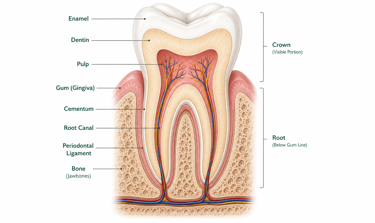
The simplest way to think about it: a tooth is a hard mineral shell (enamel) protecting a softer mineral layer (dentin), which protects a living core of nerves and blood vessels (pulp) — all anchored into the jawbone by the root and a ligament.
Enamel — the outer shell
Enamel is the visible, white part of the tooth above the gumline. It's the hardest substance in the human body — harder than bone — because its job is purely mechanical: withstand biting, chewing, and grinding forces every single day for decades.
Enamel has no nerve endings and no blood supply. That's why a cavity can grow silently through enamel with zero pain — you don't feel anything until decay reaches the layer underneath.
Dentin — the supportive layer underneath
Beneath the enamel is dentin, a yellowish layer that makes up most of the tooth's bulk. Dentin is softer than enamel and contains microscopic tubules — tiny channels that connect to the nerve in the center of the tooth.
This is why tooth sensitivity happens. When enamel wears down or gum recession exposes dentin, hot, cold, and sweet stimuli can travel through those tubules straight to the nerve, causing that sharp, sudden zing.
Pulp — the living core
At the center of every tooth is the pulp chamber, containing nerve fibers, blood vessels, and connective tissue. This is the only part of a tooth that's actually alive in the way most people think of "alive" — it can become inflamed, infected, and it can die.
When a cavity reaches the pulp, or trauma damages it, the result is the kind of deep, throbbing pain that usually sends people to the dentist immediately. A root canal procedure removes infected or dying pulp tissue while preserving the outer structure of the tooth.
Cementum and the root
Below the gumline, the outer surface of the tooth is no longer enamel — it's cementum, a thinner, softer mineralized layer that covers the root. Cementum is where the periodontal ligament attaches, anchoring the tooth into its socket.
Cementum is softer than enamel and isn't designed to be exposed to the mouth. When gum recession exposes the root surface, that area is more vulnerable to decay and sensitivity than the enamel-covered crown.
The CEJ — where crown meets root
The cementoenamel junction (CEJ) is the line where the enamel of the crown meets the cementum of the root — essentially, the tooth's "neck." In a healthy mouth, this line sits right at or just below the gumline. When gums recede, the CEJ becomes visible, which is often the first sign a patient notices that their gums are receding.
Periodontal ligament and bone — what holds it all in place
A tooth isn't fused directly to bone. Between the root and the jawbone is the periodontal ligament (PDL) — a network of collagen fibers that suspends the tooth in its socket. This ligament absorbs the shock of biting and chewing and contains nerve endings that let you sense pressure, which is part of why you can feel a popcorn kernel hiding between your teeth before you ever see it.
The bone itself, called the alveolar bone, forms the socket the tooth sits in. Gum disease that progresses untreated can destroy this bone over time — which is the actual mechanism behind teeth becoming loose or eventually being lost.
Why this matters for your dental visits
Understanding these layers explains a lot of what your dentist tells you:
- Why a small cavity needs filling now — it's still confined to enamel or outer dentin, and waiting risks it reaching the pulp
- Why a deep cavity sometimes needs a root canal instead of a filling — once decay reaches the pulp, the nerve tissue is compromised and can't heal on its own
- Why receding gums increase sensitivity — exposed cementum and dentin have a much more direct path to the nerve than enamel does
- Why X-rays matter — they're the only way to see what's happening inside the tooth and below the gumline, where the dentin, pulp, root, and bone live
The bottom line
A tooth is a layered structure built to protect a living nerve at its core. Enamel and dentin are the armor; pulp is the part that can actually hurt and heal; cementum, the ligament, and bone are what keep the whole thing anchored in place. Once you can picture these layers, almost every explanation your dentist gives you about a cavity, a root canal, or gum recession makes a lot more sense.
Read the full article
Create a free SmileSmart account to unlock every article instantly.